
5
1
9781405138994


An Atlas of Interpretative Radiographic Anatomy of the Dog and Cat / Edition 2 available in Hardcover, eBook


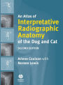
An Atlas of Interpretative Radiographic Anatomy of the Dog and Cat / Edition 2

- ISBN-10:
- 1405138998
- ISBN-13:
- 9781405138994
- Pub. Date:
- 06/03/2008
- Publisher:
- Wiley
An Atlas of Interpretative Radiographic Anatomy of the Dog and Cat / Edition 2
$315.95
315.95
In Stock

From the B&N Reads Blog