


eBook
Related collections and offers
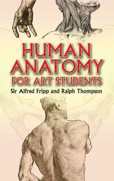
Overview
Subjects include the skeleton, the coverings and regions of the body, the upper and lower extremities, movements of the joints, the trunk, and the head and neck. Additional topics encompass expression and gesture, differences of size and proportion in the sexes, and growth, development, and measurements. More than 130 detailed figures appear throughout the text, in addition to thirty-one plates.

Product Details
| ISBN-13: | 9780486149752 |
|---|---|
| Publisher: | Dover Publications |
| Publication date: | 06/15/2012 |
| Series: | Dover Anatomy for Artists |
| Sold by: | Barnes & Noble |
| Format: | eBook |
| Pages: | 352 |
| File size: | 16 MB |
| Note: | This product may take a few minutes to download. |
Read an Excerpt
Human Anatomy for Art Students
By Alfred Fripp, Ralph Thompson
Dover Publications, Inc.
Copyright © 2006 Dover Publications, Inc.All rights reserved.
ISBN: 978-0-486-14975-2
CHAPTER 1
THE SKELETON
No bone is either quite rigid or quite straight. The elasticity (which is greater in the young) and the curves found in every bone are obviously adapted to increase its strength.
The skeleton of the adult is built up of almost rigid bones, and the length of each bone is slightly increased by a layer of less rigid cartilage, forming a kind of buffer at each end. In addition, within certain of the joints, actual cushions of fibro-cartilage intervene between the ends of the bones.
There are various forms of bone. The femur, or thigh-bone, may be taken as an example of the long bone, while the spine, wrist, and ankle are composed of short bones. The scapula, or shoulder-blade, and the frontal, or forehead bone, are good examples of the flat bone.
The stature of any individual depends chiefly upon the length of the long bones of the lower limb and of the short bones of the spinal column.
1. The long bones should be regarded as mechanical "levers"; every muscular action may be interpreted as a power or force applied to such a part of the bar or bone as to overcome a definite weight or resistance, and so to produce movement about a fixed point or fulcrum.
2. The short bones will plainly be less liable to fracture, and the multiplication of the cartilaginous and articulating surfaces will, of course, result in the better breaking of jars and increased mobility. Thus, if the spine were a single rigid long bone, its relation to the cranium would be that of the broom-handle to the broom-head, and the effect of a blow upon the other end, as when one sits down with a jerk, would be to drive the neck into the base of the skull, just as the handle of the broom is driven into the broom-head.
3. The flat bones are generally protective; thus the flat bones of the vault of the skull protect the delicate brain which lies in the cranium, and the flat bones on each side of the pelvis, known as the hip-bones or ossa innominata, afford protection to important viscera. In addition, flat bones provide an extensive attachment for strong muscles. Chewing or mastication, which is one of the most powerful and one of the most fundamentally important movements in the body, is brought about by the very strong muscles inserted into the jaw, which have an extensive origin from the flat bones of the cranium. In like manner the movements of the humerus are partly caused by muscles which arise from broad areas of the scapula.
A muscle is said to "arise or take origin" from that end which usually is fixed when the muscle acts, and its "insertion" is that end which usually moves most.
The femur is the longest bone in the body; the next longest are the bones of the leg and arm and forearm, some of the ribs, and then the clavicle.
The bones of the upper limb comprise:—
1. The clavicle, or collar-bone.
2. The scapula, or shoulder-blade.
3. The humerus, or bone of the arm.
4. The ulna, the inner bone of the forearm.
5. The radius, the outer bone of the forearm.
6. The carpals or bones of the wrist.
7. The metacarpals or bones of the palm of the hand.
8. The phalanges or bones of the digits.
1. The clavicle (Fig. 1) is situated in the front part of the thorax or chest, where the trunk merges into the neck. At its inner extremity it is joined to the sternum (breast-bone) by the sterno-clavicular joint. The inner ends of the right and left clavicles are about an inch apart. The outer extremity of the clavicle touches the acromion process of the scapula in the acromio-clavicular joint, and is situated at a somewhat higher level than the inner end (Fig. 1). This is the case even with people whose shoulders slope in a very marked degree.
The clavicle has its curves so arranged that there is a convexity forward in the inner part, for rather more than half the length of the bone, and a concavity forward in the outer part, for rather less than half. It is thicker and more prominent internally than externally: a cross section made through the internal half is triangular; through the external half it is flattened from above downwards.
In the clavicle, as in all other long bones, the degree of its roughness gives a fair indication of the muscularity of the individual.
The clavicle is a bone of high importance to the student of anatomy. It forms a very prominent landmark, easily seen in thin people. In well-covered and muscular subjects, however, it lies at the bottom of a furrow, a situation common in many other parts of the body, e.g. the external condyle of the humerus and the great trochanter of the femur, a dimple indicating the position of a bone which in the skeleton appears to be prominent.
The clavicle, unlike the other long bones, continues to increase much in length between the ages of twenty and twenty-five years, and thus produces, during this period, a great increase in the breadth of the shoulders, an increase which constitutes one of the chief characteristics distinguishing the adolescent boy from the adult man.
2. The scapula (Fig. 2) is chiefly visible upon the upper part of the back of the trunk, but two of its "processes are apparent from the front of the skeleton, namely, the acromion, which makes the point of the shoulder, and the coracoid, which is covered by thick muscles, but in the wasted subject can be seen under the skin below the outer part of the clavicle.
It is a triangular flat bone having two sur. faces:—
a. The front or ventral surface, applied to the ribs over the back of the thorax, from the second to the seventh.
b. The hinder or dorsal surface, overlaid by the muscles and skin of the back.
Three borders:—
[a. The upper.
b. The mesial, vertical, or vertebral.
c. The external, oblique, or axillary (lying in relation to the axilla or armpit).
And three angles:—
a. The superior.
b. The inferior, which is prominent in persons who are "round-shouldered."
c. The external, sometimes called the head of the scapula, which, with the head of the humerus and the connecting ligaments, forms the shoulder-joint.
The three processes of the scapula are:—
a. The spine or spinous process (Fig. 2, p. 36). This is a well-marked bony ridge which projects backwards from the dorsal surface. It begins internally at the junction of the upper and second quarter of the vertebral border, and becomes more prominent externally, where it terminates in the second process.
b. The acromion process. This is flattened and directed forwards, upwards, and outwards, to form the point of the shoulder. The acromion process has two borders, of which the inner one enters, with the external end of the clavicle, into the formation of the acromio-clavicular joint.
c. The coracoid process is curved upon itself, and tapers rapidly to its apex, which is directed forwards and outwards just below the forward concavity of the outer third of the clavicle (Fig. 1).
3. The humerus (Fig. 3), or bone of the arm proper, is described, like all the long bones, as consisting of a shaft and two extremities, upper and lower. (The extremities of the clavicle only are known as outer and inner, and the extremities of the ribs are called anterior and posterior.)
The humerus articulates above with the head of the scapula, to form the shoulder-joint, and below with the ulna and radius, where it forms the elbow-joint.
The upper extremity, or head of the humerus, forms a small segment of a large globe. It is directed upwards, inwards, and backwards, and the size of it greatly influences the prominently convex shape and outline of the shoulder. When the arm is outstretched, the head of the humerus may be felt, or even seen, in the axilla, especially in thin people.
The tuberosities of the humerus are flattened projections which are separated from the head by the anatomical neck, to which the capsule of the shoulder-joint is attached. The vertical bicipital groove divides the greater and lesser tuberosities from each other, the groove running downwards, inwards, and slightly forward, and lodging the tendon of the long head of the biceps muscle. The greater tuberosity lies outside and behind the lesser.
The shaft of the humerus begins below the tuberosities and head at a decidedly narrower part, which is known as the surgical neck on account of the frequency with which the bone is broken in this region. Below the neck the shaft becomes a little thicker, and is twisted, not bent, outwards through 38 an angle of fifteen or twenty degrees. In its lower third it is slightly flattened from before backwards, and is concave forwards.
The lower extremity of the humerus is very wide from side to side.
The condyles of the humerus lie on each side of the lower extremity. The internal, which is larger and lies at a lower level than the external, is directed chiefly inwards but also slightly backwards, while the external is directed outwards.
The condyles serve the usual purpose of bony prominences, namely, the attachment of muscles in this case, and of some of the ligaments of the neighbouring joint, the elbow. They form the lower limits of corresponding ridges, called the supra-condylar ridges, which descend on each side of the shaft of the humerus.
The trochlea is a broad, smooth articular surface at the lower end of the humerus, grooved obliquely, so that behind the groove it is directed downwards and inwards, and in front it runs upwards and outwards. The inner lip of the trochlea is more prominent than the outer, especially towards its lower part. This lip, by its large size, is responsible for the maintenance of the carrying angle (Fig. 4). The articulation of the trochlea and the ulna forms the main part of the elbow-joint (Fig. 5).
The capitellum lies between the trochlea and the external condyle and articulates with the radius in the elbow-joint. It is a rounded surface, and is not so well seen from behind as from in front.
The bones of the forearm (Fig. 6) are the radius and the ulna. The two bones lie nearly parallel to each other in the position of "attention," which is that of "supination" of the forearm; but the ulna begins higher up the limb and does not reach so far down as the radius. In pronation the radius crosses obliquely downwards and inwards over the ulna.
They taper in different directions, the ulna being larger at the elbow end and the radius at the wrist. The elbow-joint is in great part formed by the ulna, while the radius is the more important constituent of the wrist.
4. The ulna, the longer bone, lies to the inner side of the radius. The greater sigmoid cavity of the ulna is a deep hollow at its upper end, the concavity of which is directed forwards and articulates with the trochlea of the humerus. This cavity is overhung above and behind by the prominent olecranon process. The strong portion of bone known as the coronoid process projects forwards below the greater sigmoid cavity.
The lesser sigmoid cavity lies on the outer side of the upper part of the coronoid process, and receives the head of the radius.
The posterior surface of the olecranon process is triangular in shape and very easily felt, because subcutaneous. Its upper part forms the point of the elbow. The triangular area, with its apex below, is continuous with the sinuous posterior border of the shaft, which is also subcutaneous in the whole length of the bone.
The shaft tapers towards the wrist where the lower end of the ulna presents two prominences separated by a deep groove. The larger, the head of the ulna, is very obvious on the back of the wrist in the pronated position. The slenderer styloid process is detected with more difficulty by the examining finger.
Notice that the ulna tapers from elbow to wrist in about the same delicate graduation as does the undissected forearm, and that its shaft is not only concave forwards throughout its whole length, but also, when viewed from before back, it is concave outwards towards the radius.
5. The radius has a head with a saucer-like depression at its upper end. The constriction below the head is the neck, from which the shaft gradually swells out till it reaches the very large lower extremity at the wrist.
The bicipital tuberosity lies just inside and below the neck, and its posterior part gives attachment to the biceps tendon.
The shaft is convex outwards, and concave towards the ulna. At the most prominent part of the curve is a noticeable rough area, into which an important muscle known as the "pronator radii teres" is inserted.
The styloid process of the radius can be felt in the "anatomist's snuff-box" (vide p.101); it is more massive, and situated at a lower level than the corresponding process of the ulna. The back of the lower end of the radius is marked by four grooves, the deepest of which lies on the inner side of a prominent tubercle, and lodges the tendon of the extensor longus pollicis muscle.
The sigmoid cavity, lying on the inner side of the lower end of the radius, receives the head of the ulna.
The radius is joined by ligaments to the ulna. The chief of these are the collar-like orbicular ligament which surrounds the head of the radius, the triangular fibro-cartilage which unites the lower ends of the two bones, and the interosseous membrane, the fibres of which pass downwards and inwards from the shaft of the radius to that of the ulna.
This membranous ligament is a very important factor in the mechanics of the forearm. When a thrust with the hand is made, pressure is transmitted, from the object pushed to the lower end of the radius, and this bone tends to be displaced upwards. But the direction of the fibres of the interosseous membrane is such that they are at once made tight, and so they pull upon the ulna. The thrusting force is thus transferred to the ulna, distributed throughout its whole length, but changed into a pulling one, and so shock is diminished.
6. There are eight bones of the wrist or carpus (Fig. 7). They are so articulated with each other that there is a distinct general concavity of the anterior aspect of the carpus. This concavity is bounded on the inner side by the pisiform bone, the smallest of the series, and by the well-marked hook of the unciform bone, which lies more deeply and is just below the pisiform.
The corresponding eminences which bound the concavity to the outer side are the tubercle of the scaphoid and the ridge on the trapezium. The latter bone is on the outer side of a deep groove that receives the flexor carpi radialis tendon. It possesses a very special importance in that it has a small process of bone which is directed downwards and inwards and throws the thumb away from the finger. The great range of movement in the thumb is characteristic of the human hand, and is of vital importance to man in the performance of many of the finer movements which in the process of civilisation he has acquired, such as are for instance essential to the proper use of pen and pencil. This range is so free that the palmar surface of the thumb's terminal phalanx can be opposed to the palmar surface of any part of the other digits.
7. The metacarpal bones (Fig. 7, p. 43) are five, one to each digit. Their posterior surfaces form longitudinal ridges on the back of the hand, more obvious in the aged or emaciated, and their heads form the first or proximal set of knuckles.
8. The phalanges (Fig. 7) are fourteen in number —three to each finger, but only two to the thumb. They diminish in size from above downwards, and are described as long bones, in that each has a shaft and two extremities. The heads of the proximal and middle phalanges form the middle and distal rows of knuckles.
The bones of the lower limb comprise:—
1. The os innominatum, or hip-bone. The bony basin of the pelvis is formed of the two ossa innominata together with the sacrum and coccyx, which are the two lowest bones of the spinal column.
2. The femur, or thigh-bone.
3. The patella, or knee-cap.
4. The tibia, or large bone of the leg.
5. The fibula, or small bone of the leg.
6. The tarsals.
7. The metatarsals.
8. The phalanges.
1. The hip-bone belongs to the class of flat bones. Its functions are:—
To give attachment to the strong muscles which maintain the erect position (Figs. 8 and 9, p. 45).
To support the weight of the head, trunk, and upper limbs, and to transmit this weight to the lower limb.
To protect the important viscera which lie in the lower part of the abdominal cavity and in the pelvis.
The relation of the os innominatum to the sacrum in the constitution of the pelvic cavity will be described later.
(Continues...)
Excerpted from Human Anatomy for Art Students by Alfred Fripp, Ralph Thompson. Copyright © 2006 Dover Publications, Inc.. Excerpted by permission of Dover Publications, Inc..
All rights reserved. No part of this excerpt may be reproduced or reprinted without permission in writing from the publisher.
Excerpts are provided by Dial-A-Book Inc. solely for the personal use of visitors to this web site.
Table of Contents
IntroductionI. The Skeleton
II. The Coverings of the Body
III. The Regions of the Body
IV. The Upper Extremity
V. The Lower Extremity
VI. Movements of the Joints of the Upper Extremity
VII. Movements of the Joints of the Lower Extremity
VIII. The Trunk
IX. The Head and Neck
X. Expression and Gesture
XI. Differences of Size and Proportion in the Sexes
XII. Growth, Development, and Measurements
Appendix on Comparative Anatomy
Index